Table of Contents
Popliteal Fossa Introduction
Popliteal fossa(Latin hamstring of the knee) is a shallow diamond-shaped depression felt best at the back of the knee joint when the joint is semi-flexed. It corresponds to the cubital fossa of the forearm.
Surface Landmarks
- Lateral and medial condyles of the femur and tibia can be identified easily on the sides and front of the knee.
- The Head of the fibula is a bony prominence situated just below the posterolateral aspect of the lateral condyle of the tibia.
- The common peroneal nerve can be palpated against the posterolateral aspect of the neck of the fibula, medial to the tendon of the biceps femoris, by moving the finger from below upwards.
- The fibular collateral ligament of the knee joint is felt like a rounded cord just above the head of the fibula in a flexed knee.
- When the knee is flexed against resistance, the hamstrings can be seen and palpated easily right up to their insertion. Medially, the rounded tendon of the semitendinosus lies superficial to the flat tendon of semimembranosus. In front of these tendons, there is a groove bounded anteriorly by the tendon of the adductor Magnus. Laterally, there is the tendon of the biceps femoris. In front of this tendon, there is a shallow groove bounded anteriorly by the iliotibial tract.
- Pulsations of the popliteal artery can be felt in the middle of the popliteal fossa by applying deep pressure.
- In the lower part of the popliteal fossa, two heads of the gastrocnemius form rounded cushions that merge inferiorly into the calf.
Dissection of Popliteal Fossa
- Make a horizontal incision across the back of the thigh at its junction of upper two-thirds with lower one-third and another horizontal incision at the back of the leg at its junction of upper one-third and lower two-thirds.
- Draw a vertical incision joining the midpoints of the two horizontal incisions made. Reflect the skin and fascia on either side.
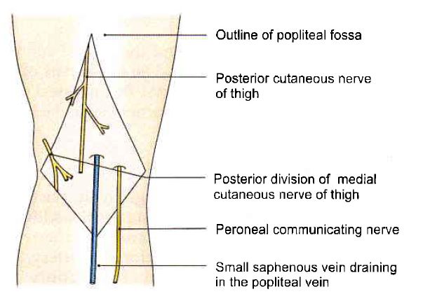
- Find the cutaneous nerves, e.g. posterior cutaneous nerve of thigh, posterior division of medial cutaneous nerve, sural communicating nerve, and short saphenous vein. Cut and clean the deep fascia.
- Identify the boundaries and contents of the fossa.
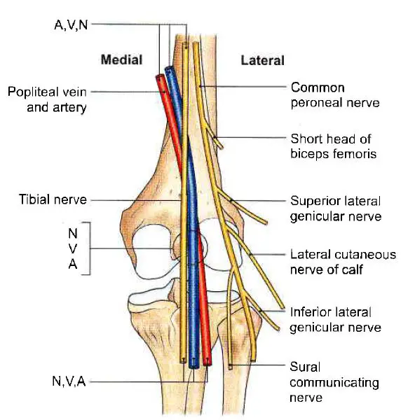
- Trace the tibial nerve as it courses through the center of the popliteal fossa. Its three delicate articular branches are given off in the upper part of the fossa, cutaneous branch in the middle part, and muscular branches in the lower part of the fossa.
- The common peroneal nerve is lying just medial to the tendon of the biceps femoris muscle. Trace its branches.
- The popliteal vein is deep to the tibial nerve and the popliteal artery is the deepest as seen from the back. Trace all the muscular, cutaneous, genicular, and terminal branches of the popliteal artery.
Specific Location
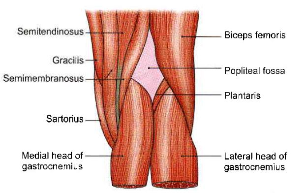
The popliteal fossa is a diamond-shaped depression lying behind the knee joint, the lower part of the femur, and the upper part of the tibia.
Boundaries of Popliteal Fossa
Superolaterally: The biceps femoris.
Superomedially: The semitendinosus and the semimembranosus, supplemented by the gracilis, the sartorius, and the adductor Magnus.
Inferolaterally: The gastrocnemius lateral head is substituted by the plantaris.
Inferomedially: Gastrocnemius(medial head)
The roof of the popliteal fossa is formed by the deep fascia or popliteal fascia.
The superficial fascia over the roof contains;
- The small saphenous vein and cutaneous nerves.
- Three cutaneous nerves, namely, the branches and terminal part of the posterior cutaneous nerve of the thigh, the posterior division of the medial cutaneous nerve of the thigh, and the peroneal or sural communicating nerve.
The popliteal fossa’s floor is developed from above downwards by:
- The femur’s popliteal surface.
- The knee joint capsule and the oblique popliteal ligament.
- The popliteal fascia is a tight fascia that covers the popliteus muscle.
Contents of the Popliteal Fossa
- The popliteal artery and its branches.
- The popliteal vein and its tributaries.
- The tibial nerve and its branches.
- The common peroneal nerve and its branches.
- The posterior cutaneous nerve of the thigh.
- The genicular branch of the obturator nerve.
- The popliteal lymph nodes.
The popliteal vessels and tibial nerve cross the fossa vertically, one on top of the other. The tibial nerve is the most superficial, followed by the popliteal vein, which is deep or anterior to the tibial nerve, and finally, the popliteal artery, which is deepest of all. The vein and nerve both cross the artery posteriorly.
These systems are arranged in the following order;
- In the upper part of the fossa, from medial to lateral side: artery, vein, and nerve (A, V, N).
- In the middle part, from behind forwards: nerve, vein, and artery (N, V, A).
- In the lower part, from medial to lateral side: nerve, vein, and artery (N, V, A).
The typical peroneal nerve runs obliquely through the fossa from the superior angle to the lateral angle, along the medial border of the biceps femoris, and in the same superficial plane as the tibial nerve.
The Popliteal Artery
Read this article to study all about the Popliteal Artery.
The Popliteal Vein
Read this article to study all about the Popliteal Vein.
The Tibial Nerve
Read this article to study all about Tibial Nerve.
The Common Peroneal Nerve
Read this article to study all about Common Peroneal Nerve.
Posterior Cutaneous Nerve of the Thigh
It is a component of the popliteal fossa’s upper half. It pierces the deep fascia near the center of the fossa and supplies the skin all the way up to the middle of the back of the leg.
Genicular Branch of Obturator Nerve
This is the extension of the obturator nerve’s posterior division. It passes on the popliteal artery’s posterior surface, pierces the oblique popliteal ligament, and supplies the knee capsule.
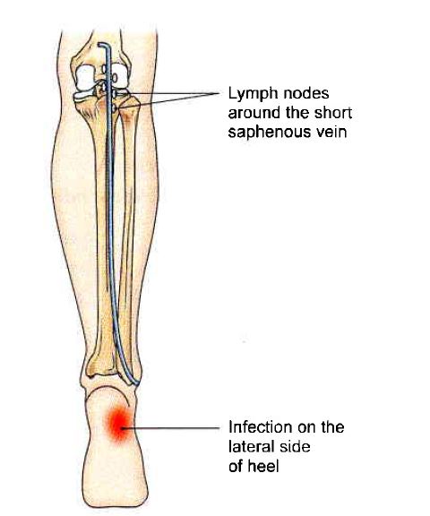
Popliteal Lymph Nodes
These are located deep to the deep fascia at the end of the narrow saphenous vein. They obtain afferents from the lateral portion of the sole, the back of the leg (both superficial and deep), and the knee joint. Efferents terminate in deep inguinal lymph nodes.
Infection on the lateral side of the sole/foot causes enlargement of the popliteal lymph nodes. These are found in the saphenous vein, which is a small vein. The saphenous vein is a short vein that pierces deep fascia to flow into the popliteal vein.
Anastomoses Around The Knee Joint
- Anastomoses around the knee joint are a complex arterial network that runs around the patella, the lower end of the femur, and the upper end of the tibia.
- The network is divided into two parts: superficial and deep. The superficial component is located in the superficial fascia surrounding the patella and in fat behind the ligamentum patellae. The deep component is found all over the articular surfaces of the femur and tibia.
- It is formed:
Medially, and above the condyle;
- The descending genicular branch of the femoral and its saphenous branch.
- The superior medial genicular.
Medially and below the condyle;
- Saphenous branch of descending genicular
- The inferior medial genicular.
Laterally, and above the condyle;
- The descending branch of the lateral circumflex femoral.
- The superior lateral genicular.
Laterally and below the condyle;
- The inferior lateral genicular.
- Anterior tibial recurrent.
- Posterior tibial recurrent from anterior tibial.
- Circumflex fibular from posterior tibial The medial and lateral arteries form longitudinal anastomoses on each side of the patella. The longitudinal anastomoses are interconnected to form transverse anastomoses just above and below the patella and above the tibial tuberosity.
- The anastomoses supply the bones that make up the knee joint, as well as the capsule and synovial membrane.
Mnemonics for the Popliteal Fossa
The upper part, from the medial to the lateral side
- AVN
A-Popliteal artery
V-Popliteal vein
N—Tibial nerve
The middle part, from behind forwards
- NVA
N—Tibial nerve
V-Popliteal vein
A—Popliteal artery
The lower part, from the medial to the lateral side
- NVA
N-Tibial nerve
V-Popliteal vein
A—Popliteal artery
Read Cubital Fossa
Last Updated on February 23, 2022 by Learn From Doctor Team


