Table of Contents
Venous Bleeding Overview
In case of venous bleeding blood flow comes out from a vein. The function of the vein is to transport deoxygenated blood to the heart. There are three types of bleeding that occurs;
- Arterial bleeding: It occurs when the blood flow comes out from the artery.
- Venous bleeding
- Capillary bleeding: It occurs when blood flow comes out from very small vessels which connect the artery and veins.
The most severe and dangerous type of bleeding is arterial bleeding. But in certain circumstances, venous bleeding can be also dangerous and fatal. There are some signs and features by which we can detect the bleeding types and severity. Let’s discuss them.
Signs of Bleeding Vein
When a vein is cut or ripped, the blood that comes out is dark red or bluish. This is due to a lack of oxygen.
Furthermore, because venous blood is going to the heart, it is subjected to less pressure than arterial blood. It will “ooze” like a thick liquid, slowly draining from the body. Blood can gush out if the affected vein is wide or big.
Wounds that may cause bleeding vein;
- Punctured wound
- Laceration
- Amputation
The First Step to Manage the Bleeding from the Vein
In general, all forms of bleeding necessitate the same treatment. The aim is to stop the bleeding, avoid blood loss, and repair the underlying vein rupture or laceration.
Here’s what you can do if you meet someone who has a bleeding vein:
- To cover yourself, put on a pair of latex gloves. Wrap your hands in a plastic bag or layers of clean fabric if you don’t have gloves.
- Locate the wound. Remove or cut the person’s clothes if necessary to reveal the wound properly.
- Elevate the wound above the person’s heart if necessary.
- Apply clean gauze or fabric, such as a handkerchief, to the wound. If you don’t have these things, use your hand instead.
- For 5 minutes, apply consistent, strong pressure. If the wound is thin, use your fingers to close it. Use your palm if the wound is big.
- If the bleeding continues for more than 10 minutes, put another cloth on top.
- Firmer pressure should be applied over a larger region. Avoid removing the first layer of soaked fabric because it can disrupt clotting.
- If the bleeding does not stop, if there is a lot of bleeding, or if the person loses consciousness, dial 911.
Venous bleeding is usually less difficult to control than arterial bleeding. However, if the vein is very deep or wide, it might be difficult to stop the bleeding.
Emergency Situations
Most of the time bleeding from the vein isn’t serious. It can be stopped by the above-mentioned first steps But if the vein is deep and wide then sometimes the bleeding cannot be stopped and needed more emergency support.
Signs of emergency
- After several minutes of applying pressure, the bleeding does not stop or tends to stop.
- The bleeding is spat out rapidly, indicating arterial bleeding.
- The wound is big, deep, or embedded with an object.
- The wound reveals the bone.
- The wound affects the eyes or the abdomen.
- The wound is in the chest or neck which makes breathing difficult.
- The injury was caused by a car crash.
- The person exhibits signs of shock.
Venous Injury Inspection
Venous Drainage of Upper Limb
The upper limb venous system drains deoxygenated blood from the arm, forearm, and hand. It is divided into two parts:
- The superficial system
- The deep system.
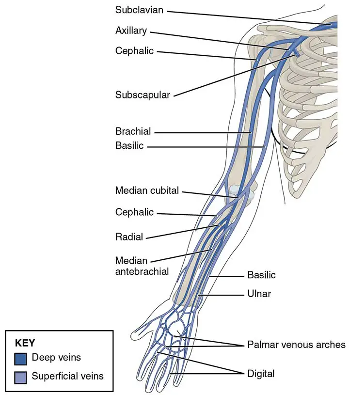
The Superficial Veins
The cephalic and basilic veins are the main superficial veins of the upper extremity. They are found in the upper limb’s subcutaneous tissue.
The Basilic Vein
The basilic vein arises from the hand’s dorsal venous network and ascends the medial side of the upper limb.
The vein travels deep into the arm at the teres major border. The axillary vein is formed as it joins the brachial veins of the deep venous system.
The Cephalic Vein
The dorsal venous network of the hand also gives rise to the cephalic vein. It ascends the anterolateral part of the upper limb and passes anteriorly at the elbow.
The cephalic vein passes between the deltoid and pectoralis major muscles at the shoulder (known as the deltopectoral groove) and reaches the axilla through the clavipectoral triangle. The cephalic vein empties into the axillary vein inside the axilla.
The median cubital vein connects the cephalic and basilic veins at the elbow joint.
The Deep Veins
The upper limb’s deep venous system is located beneath the deep fascia. It is made up of paired veins that run alongside and on either side of an artery. The deep veins in the upper extremity are named for the artery that they surround.
The brachial veins are the largest and are located on either side of the brachial artery. The brachial artery pulsations aid in venous return. Vena comitantes are veins that are structured in this manner.
Perforating veins link the two systems by running between the deep and superficial veins of the upper limb.
Venous Drainage of Lower Limb
Lower limb veins drain deoxygenated blood from thigh, gluteal region, leg and foot and return it to the heart. They are classified into two types:
- Deep: Deep veins run alongside the main arteries under the deep fascia of the lower limb.
- Superficial: In the subcutaneous tissue, superficial veins can be located. They finally empty into deep veins.
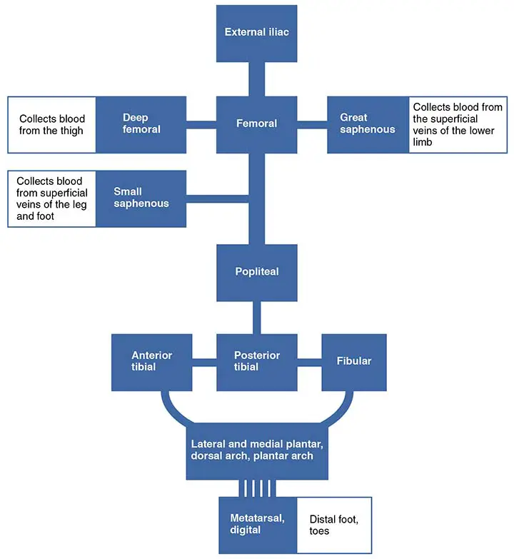
Deep Veins of Lower Limb
The deep venous drainage system of the lower limb is situated under the lower limb’s deep fascia. Deep veins typically complement and share the name of the main arteries in the lower limb. The artery and vein are often found inside the same vascular sheath, allowing the arterial pulsations to aid venous return.
The Leg and Foot
The dorsal venous arch is the largest venous structure of the foot, and it flows primarily into the superficial veins. The anterior tibial vein is formed as veins from the arch reach deep into the leg.
The medial and lateral plantar veins appear on the plantar side of the foot. The posterior tibial and fibular veins are formed by the union of these veins. The posterior tibial vein joins the posterior tibial artery at the medial malleolus and enters the leg posteriorly.
The anterior tibial, posterior tibial, and fibular veins join to form the popliteal vein on the back of the knee. The adductor canal is where the popliteal vein reaches the leg.
The Thigh
The popliteal vein becomes the femoral vein until it enters the thigh. It runs anteriorly alongside the femoral artery.
The other major venous structure in the thigh is the deep vein (profunda femoris vein). It drains deoxygenated blood from the thigh muscles by perforating veins. It then empties into the femoral vein’s distal portion.
The femoral vein exits the thigh by passing under the inguinal ligament, where it becomes the external iliac vein.
The Gluteal Region
The inferior and superior gluteal veins drain the gluteal area. These veins drain into the internal iliac vein.
Superficial Veins of Lower Limb
The superficial veins of the lower limb flow under the skin. The great saphenous vein and the small saphenous vein are the two main superficial veins.
The Great Saphenous Vein
The dorsal venous arch of the foot and the dorsal vein of the great toe come together to form the great saphenous vein. It runs up the medial side of the thigh, anteriorly to the medial malleolus at the ankle, and posterior aspect to the medial condyle at the knee.
The vein receives tributaries from other small superficial veins as it travels up the leg. The great saphenous vein drains into the femoral vein directly below the inguinal ligament.
The great saphenous vein can be extracted surgically and used as a vessel in coronary artery bypasses.
The Small Saphenous Vein
The dorsal venous arch of the foot and the dorsal vein of the little toe combine to form the narrow saphenous vein. It travels up the back of the leg, past the lateral malleolus, and along the lateral edge of the calcaneal tendon. It runs here between two heads of the gastrocnemius muscle before emptying into the popliteal vein in the popliteal fossa.
Few Last Words
When a vein is ripped or cut, it causes venous bleeding. The blood will appear dark red and will ooze out of the body steadily and painfully. It will not erupt like arterial blood.
While venous bleeding appears to be less severe than arterial bleeding, it can be just as dangerous. It is important to apply firm pressure to the wound as soon as possible to stop the bleeding.
Last Updated on February 23, 2022 by Learn From Doctor Team


