Table of Contents
Common Peroneal Nerve
Peroneal Nerve Root value: Dorsal divisions of ventral rami of L4, L5, S1, S2. The peroneal nerve is the sciatic nerve’s smaller terminal branch. The tibial nerve is in the same superficial plane as it. It runs along the medial border of the biceps femoris from the superior angle of the fossa to the lateral angle. It winds along the posterolateral aspect of the fibula’s neck, pierces the peroneus longus, and splits into the superficial and deep peroneal nerves as it continues downwards and forwards.
Read Popliteal Fossa
Branches
Cutaneous branches are two types
- The lateral cutaneous nerve of the calf descends to supply the skin of the upper two-thirds of the lateral side of the leg.
- Sural communicating nerve arises in the upper part of the fossa. It runs on the posterolateral aspect of the calf and joins the sural nerve.
Articular branches
- The superior lateral genicular nerve accompanies the artery of the same name and lies above the lateral femoral condyle.
- The inferior lateral genicular nerve also runs with the artery of the same name to the lateral aspect of the knee joint above the head of the fibula.
- Recurrent genicular nerve arises where common peroneal nerve divides into superficial and deep. peroneal nerves. It ascends anterior to the knee joint and supplies tibialis anterior muscle in addition to the knee joint.
Read The Tibial Nerve
Muscular branches
- They do not arise from this nerve. However, it may give a branch to the short head of the biceps femoris.
Surface Marking
It is marked by connecting the two points mentioned below.
- The first point is located at the apex of the popliteal fossa.
- The second point is on the back of the fibula’s spine.
The nerve curves forward at the lower end and ends deep to the upper fibers of the peroneus longus.
Clinical Anatomy of Common Peroneal Nerve
The common peroneal nerve can be palpated against the fibula’s posterolateral neck. It could be hurt in this place. It is the most often damaged nerve in the lower extremity. This nerve is not well-protected. It can become entangled between the peroneus longus attachments to the head and shaft of the fibula. People experience “foot drop,” which is usually painless. There is a lack of ankle dorsiflexion and eversion of the foot. Inversion and plantar flexion are common, and the ankle jerk is still present.
Read Popliteal Artery
Superficial Peroneal Nerve
Dissection
Examine the common peroneal nerve in relation to the fibula’s neck.
The superficial peroneal nerve, one of the terminal branches of the common peroneal nerve, supplies both the peroneus longus and brevis muscles and becomes cutaneous in the lower one-third of the leg to supply almost all of the dorsum of the foot.
Overview of Superficial Peroneal Nerve
The main nerve of the lateral compartment of the leg is the superficial peroneal nerve.
Origin
It is a smaller terminal branch of the common peroneal nerve.
Course
The nerve starts on the lateral side of the fibula’s neck, passes through the peroneal muscles, and becomes superficial at the upper two-thirds and lower one-third of the leg.
Relation
It starts on the lateral side of the fibula’s neck, under the upper fibers of the peroneus longus.
It descends through the substance of the peroneus longus in the upper one-third of the leg.
In the middle one-third, it first descends for a short distance between the peroneus longus and brevis muscles, then descends in a groove between the peroneus brevis and the extensor digitorum longus under cover of deep fascia.
It pierces the deep fascia to become superficial at the junction of the upper two-thirds and lower one-third of the leg. It splits into two branches, one medial and one lateral, which descend into the foot.
Read Popliteal Vein
Branches along with distribution
Muscular Branches
- The Peroneus longus
- The peroneus brevis
Cutaneous Branches
Except for the territories supplied by the saphenous, sural, deep peroneal nerves and plantar nerves, the superficial peroneal nerve supplies the lower one-third of the lateral side of the leg and the greater part of the dorsum of the foot through its terminal branches.
The Medial Branch
The medial branch crosses the ankle and splits into two dorsal digital nerves, one for the medial side of the big toe and the other for the adjoining sides of the second and third toes.
The Lateral Branch
The lateral branch also splits into two dorsal digital nerves for the third and fourth toes, as well as the fourth and fifth toes.
The Communicating Branches
Branches communicating. The saphenous and deep peroneal nerves associate with the medial branch, while the sural nerve communicates with the lateral branch.
Nerve supply and actions of the peroneal muscles Nerve supply
| Muscle | Nerve Supply | Action |
| Peroneus Longus | Superficial Peroneal Nerve |
|
| Peroneus Brevis | Superficial peroneal Nerve | Evertor of the foot |
Surface Marking
It is marked by connecting the two points mentioned below.
- The first point is on the lateral aspect of the fibula’s neck.
- Second point: At the junction of the upper two-thirds and lower one-third of the leg, on the anterior border of the peroneus longus.
The nerve pierces the deep fascia at the lower end and splits into medial and lateral branches.
Clinical Anatomy of Superficial Peroneal Nerve
The superficial peroneal nerve supplies both peronei before splitting into medial and lateral cutaneous branches that supply the dorsum of the foot. As it penetrates the deep fascia of the leg, the superficial peroneal nerve can become entrapped. It is also thought to be involved in lateral compartment syndrome. Its paralysis results in a lack of eversion of the foot at the subtalar joint.
Deep Peroneal Nerve
Overview of Deep Peroneal Nerve
The deep peroneal nerve is a nerve that runs through the anterior compartment of the leg and the dorsum of the foot. It refers to the forearm’s posterior interosseous nerve. This is one of two terminal branches of the common peroneal nerve that emerge between the fibula’s neck and the peroneus longus muscle.
The Relation and Course
The deep peroneal nerve starts on the lateral side of the fibula’s spine, under the cover of the peroneus longus upper fibers By penetrating the anterior intermuscular septum, it enters the anterior compartment of the leg. It then pierces the extensor digitorum longus and arrives at the anterior tibial vessels.
It is associated with the anterior tibial artery in the leg and has identical relationships. In the upper and lower thirds of the leg, the nerve is lateral to the artery, and in the middle one-third, it is anterior to the artery.
The nerve divides into lateral and medial terminal branches on the dorsum of the foot, close to the ankle joint.
The lateral terminal branch branches laterally and terminates in a pseudoganglion deep to the extensor digitorum brevis. The extensor digitorum brevis and the tarsal joints are supplied by branches that originate from the pseudoganglion.
The medial terminal branch terminates by providing the skin adjacent to the first interdigital cleft and the big toe’s proximal joints.
Branches Along With Distribution
Muscular Branches
Muscular branches supply the muscles mentioned below.
- Muscles of the anterior compartment of the leg.
These are;
- Tibialis anterior
- Extensor hallucis longus
- Extensor digitorum longus
- Peroneus tertius.
- The extensor digitorum brevis: The lateral terminal branch of the deep peroneal nerve supplies the extensor digitorum brevis (on the dorsum of the foot).
Cutaneous Branches
The dorsal digital nerves for the big and second toes are formed by the lateral terminal branch of the deep peroneal nerve.
Articular Branches
- Ankle joint
- Tarsal joints
- Tarsometatarsal joint
- The metatarsophalangeal joint of the big toe.
Surface Marking of Deep Peroneal Nerve
It is marked by connecting the two points mentioned below.
- The first point is on the lateral aspect of the fibula’s neck.
- Second point: halfway between the two malleoli in front of the ankle.
- The third point is the first interosseous space.
In its upper and lower thirds, the nerve is lateral to the anterior tibial artery but anterior to the artery in its middle part.
Clinical Anatomy of the Peroneal Nerve
The popular peroneal nerve is the most commonly paralyzed nerve. This is injured as a result of a fibula neck fracture, a lathi injury on the lateral side of the knee joint, or a plaster on the leg. In the last instance, the nerve is compressed between the tough plaster and the fibula’s neck. Cotton must be positioned on the upper lateral side of the leg to avoid this.
The following are the consequences of injury: Motor loss in the foot’s dorsiflexor and evertors.
The standard foot position is “foot drop,” with sensory loss to the back of the leg, the lateral side of the leg, and the majority of the dorsum of the foot.
Articular loss on the lateral side of the knee joint.
The loss of dorsiflexion of the foot is caused by the paralysis of muscles in the anterior compartment of the leg. As a consequence, the foot is flexed in the plantar flexion position. The condition is known as “foot drop.”
Peroneal Nerve Injury
The sciatic nerve branches into the common peroneal nerve, which provides sensation to the front and sides of the legs as well as the tops of the feet. This nerve also controls the muscles in the leg that raise the ankle and toes. Numbness, tingling, discomfort, fatigue, and a gait problem known as foot drop can result from peroneal nerve injuries.
Causes
- Dislocation of knee
- Lower limb fracture
- Knee fracture
- Surgery on the knee or hip
- Compression on the peroneal nerve
There are also some neurological causes;
- Multiple sclerosis
- Parkinson’s disease
- Herniated lumbar disk
Symptoms of Peroneal nerve injury
- Inability to raise the ankle or point the toes upward (dorsiflexion)
- Pain, numbness, or weakness in the shin or top of the foot
- Inability to move the foot
- When the leg swings forward, the knee is lifted higher than normal to remove the foot from the ground (also called steppage or foot drop gait)
Diagnosis
- Electromyography,
- Nerve conduction study,
- CT scan
- Ultrasound
- MRI
Treatment
- Decompression surgery
- The affected nerve repair
- The affected nerve grafting
- The affected nerve transfer
- The affected tendon transfer

Summery
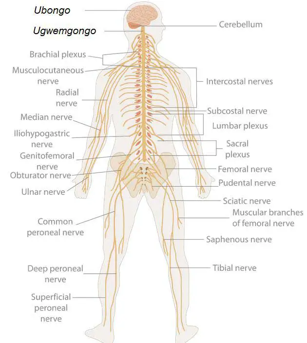
The peroneal nerve(common peroneal nerve) is the smaller terminal branch of the sciatic nerve. Its root value is the dorsal division of ventral rami of L4, L5, S1, S2 segments of the spinal cord.
Beginning: It begins in the back of the thigh as a smaller subdivision of the sciatic nerve.
Course: It lies in the upper lateral part of the popliteal fossa, along the medial border of the biceps femoris muscle. It turns around the lateral surface of the fibula. Then it lies in the substance of the peroneus longus muscle.
Branches of Common Peroneal Nerve in Popliteal Fossa
| Muscular Branch |
|
| Cutaneous and Vascular Branches |
|
| Articular Branches |
|
| Terminal Branches |
|
Termination: Ends by dividing into two terminal branches, i.e. superficial peroneal and deep peroneal nerves.
Last Updated on February 23, 2022 by Learn From Doctor Team


