Table of Contents
The Basilic Vein Beginning, Course, and Termination
The basilic vein is the upper limb’s postaxial vein (as compared to the lower limb’s short saphenous vein). It starts at the medial end of the dorsal venous arch.
It runs upwards;
- Along the back of the forearm’s medial border.
- Then winds round the medial border near the elbow,
- Continues upwards in front of the elbow (medial epicondyle) and along the medial margin of the biceps brachii up to the center of the arm, where it joins the biceps brachii.
- Where it pierces the deep fascia and runs along the medial side of the brachial artery to the lower border of the teres major, where it merges with the axillary vein, and also where it pierces the deep fascia.
- It runs along the medial side of the brachial artery until it reaches the lower boundary of the teres major, where it becomes (named as) the axillary vein.
The median cubital vein joins it about 2.5 cm above the medial epicondyle of the humerus.
It is accompanied by the posterior branch of the forearm’s medial cutaneous nerve and the terminal part of the ulnar nerve’s dorsal branch.
Read The Cephalic Vein
Detailed Anatomy
Origin
The basilic vein developed from the ulnar side of the dorsum of the hand’s superficial venous network.
Course
The basilic vein moves up posteromedially in the forearm from the ulnar aspect of the superficial venous network to the anterior elbow area, where it passes anterior to the medial epicondyle of the humerus. It continues to rise anteromedially in the brachium until it penetrates the brachial fascia at the basilic hiatus, halfway between the elbow and axilla. The basilic vein then runs medial to the brachial artery, joining the brachial veins in the axilla to form the axillary vein.
Tributaries
The upper limb’s superficial veins are extremely variable, with several superficial tributaries draining into them.
Read The Cubital Fossa
The basilic vein has two such variable tributaries;
The Median Cubital Vein
- In the anterior part of the cubital fossa, it links the basilic vein to the cephalic vein.
- It is a frequent site for venipuncture.
The Median Antebrachial Vein
- The palmar venous plexus on the palm drains the palmar digital veins. It runs anteriorly in the forearm and drains the anterior wrist and forearm subcutaneous tissue.
- It meets the basilic or median cubital veins.
- In some cases, the median antebrachial vein divides at the elbow into the median cephalic and median basilic veins, which flow into the cephalic and basilic veins, respectively.
Drainage
The basilic vein drains the medial side of the dorsum of the hand’s superficial venous network, which drains blood from the palm The basilic vein drains the medial aspect of the upper limb through numerous superficial veins as it ascends in the forearm and arm.
Termination
At the inferior border of the teres major muscle, the basilic vein joins the paired brachial veins to establish the axillary vein.
Read The Axillary Vein
Embryology
The venous system of the upper limb develops from the embryo’s anterior cardinal veins, which drain the cephalic half of the embryo properly. The remaining embryo is drained by the posterior cardinal veins. The cardinal veins form a symmetrical system throughout the fourth week. The presence of multiple anastomoses resulting in blood being channeled to the right side promotes the return of blood to the right atrium for further circulation defines the formation of the vena cava system.
The left brachiocephalic vein grows from the anastomosis between the anterior cardinal veins. The overwhelming majority of blood from the left side of the head and upper extremity is then directed to the right. The venous system in adults is much more variable than the arterial system. Individual sections emerge from various parts of the embryonic venous system during development via transformative processes.
Clinical Anatomy
Venepuncture
Venepuncture is the procedure for achieving intravenous entry, most usually for blood sampling. A hollow needle is inserted into a superficial vein through the skin (typically in the cubital fossa of the forearm). The blood is then drawn into evacuated tubes.
Varicose Vein
Read this article to read about the varicose vein.
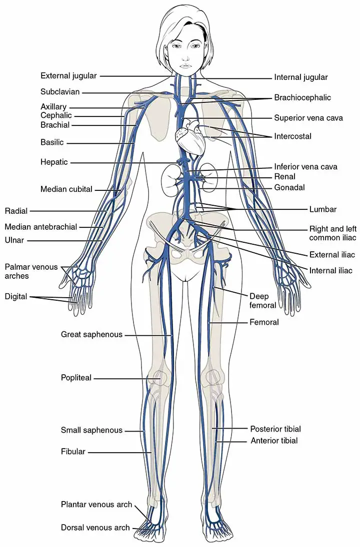
To understand the anatomy of basilic veins anatomy properly, one should study the vascular anatomy of the upper limb first.
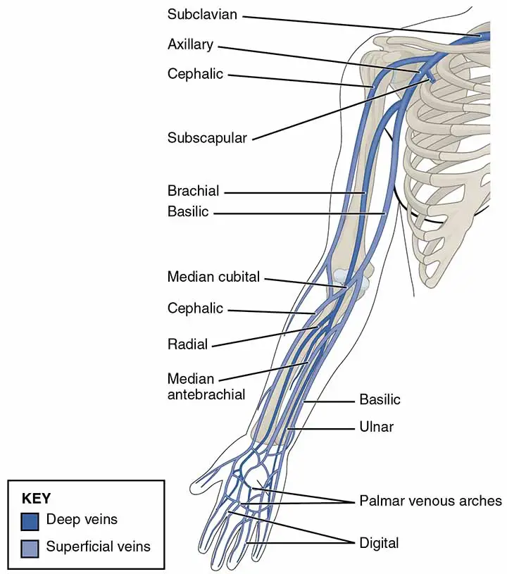
Veins of the Upper Limb
The upper limb is drained by superficial and deep vessel groups.
The superficial group begins on the back of the hand as an irregular dorsal arch. The cephalic vein starts at the radial extremity of the arch and ascends through the superficial fascia along the lateral aspect of the arm to join the deltopectoral groove. It crosses the axillary vein near the tip of the coracoid after piercing the deep fascia above the superior margin of the pectoralis minor. The basilic vein drains the ulnar end of the arch, travels along the medial aspect of the forearm, pierces the deep fascia at the elbow, and joins the brachial artery’s venae comitantes to form the axillary vein. On the flexor aspect of the elbow, the prominent median cubital vein connects the cephalic and basilic veins. It receives a number of tributaries from the flexor part of the forearm and gives off the deep median vein, which pierces the fascial roof of the antecubital fossa and joins the brachial artery’s venae comitantes.
The deep group of veins drains the tissues under the upper limb’s deep fascia and is connected to the superficial system by perforating veins. The deep veins normally follow the arteries as venae comitantes, eventually becoming the axillary, and then the subclavian, vein.
The subclavian vein is the axillary vein’s central continuation.
It begins at the inferior margin of the first rib and crosses it. The vein runs deep to the clavicular head of the pectoralis major and then to the subclavius, which acts as a barrier between it and the overlying medial part of the clavicle, before terminating at the insertion of the sternocleidomastoid.
Often only the vein’s superior margins rise above the medial end of the clavicle; other times, the entire vein does. The phrenic nerve and the inferior portion of the anterior scalenus are located posteriorly.
At the medial boundary of the scalenus anterior, behind the sternoclavicular joint, the subclavian vein joins the internal jugular vein to form the brachiocephalic vein.
Typically, a pair of valves are located within 2 cm of this intersection. The external jugular, dorsal scapular, and, on occasion, anterior jugular veins are tributaries of the subclavian vein. A forced expiration (Valsalva) maneuver demonstrates the trifurcation of the subclavian, external, and internal jugular veins, as the distended veins rise over the sternoclavicular joint, filling the fossa above the jugular notch of the sternum. The main lymphatic vessels connect to the subclavian veins at or around this junction with the internal jugular vein. The thoracic duct enters the left subclavian vein, while the right lymphatic duct enters the right subclavian vein. After wounding the subclavian, external, or internal jugular veins, there is a chance of air embolism.
Summary of the Basilic Vein
The basilic vein develops medially in the hand’s dorsal venous network. It ascends posteromedially in the forearm, inclining forward to the anterior surface distal to the elbow, where it joins the median cubital vein. It then ascends superficially between the biceps and the pronator teres, and is crossed by filaments of the forearm’s medial cutaneous nerve that travel both superficially and deeply to the vein. From the lower boundary of the teres major, the basilic vein continues as the axillary vein.
Last Updated on February 23, 2022 by Learn From Doctor Team


