Table of Contents
Parietal Bone Overview
A significant portion of the roof and sides of the skull vault are formed by two parietal bones. Each bone has an approximately quadrilateral shape with its convexity pointing outwards.
Determination of Side
- The outer surface is smooth and convex, while the inner surface is concave and has vascular markings.
- The anteroinferior angle is pointed, and a groove for the anterior division of the middle meningeal artery can be seen.
Features of the Parietal Bone
The parietal bone has two surfaces with four borders and four angles.
Surfaces
- Outer convex surface
- Inner concave surface
Borders
- Superior or sagittal border
- Inferior or squamosal border
- Anterior or frontal border
- Posterior or occipital border
Angels
- Anterosuperior or frontal angel
- Anteroinferior or sphenoidal angel
- Posterosuperior or occipital angel
- Posteroinferior or mastoid angel
Fontanelle
- One anterior fontanelle, which closes at 18 months.
- One posterior fontanelle, which closes at 3 months
- Two anterolateral or sphenoidal fontanelles, which close at 3 months.
- Two posterolateral or mastoid fontanelles which close at 12 months of life.
Anatomical Position
The side to which the bone belongs is determined by the longest serrated border above towards the median plane, the extending anteroinferior (sphenoidal) angle downward and forward, and the convex external surface.
Deep Anatomy of the Parietal Bone
The parietal bones are a pair of quadrilateral curved plates of flat bones that form the majority of the roof and sides of the skull.
Each bone has external and internal surfaces, as well as four borders and four angles.
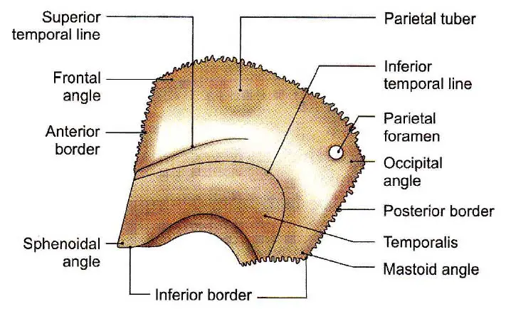
The External Surface
It is convex and smooth, with an elevation near the center called the parietal tuber or tuberosity. The superior and inferior temporal arches stretch across the bone just below the tuber with upward convexity.
The superior temporal line connects to the temporal fascia and the lateral portion of the epicranial aponeurosis, and it continues further downward superficial to the temporal fascia to connect to the upper boundary of the zygomatic arch as well as the temporal fascia. The inferior temporal line and the region of bone under it forms the majority of the temporal fossa floor and give rise to the temporalis muscle. The bone is shielded by the epicranial aponeurosis above the superior temporal line; close to the posterior part of the superior border, the external surface normally has a parietal foramen that transmits an emissary vein linking the superior sagittal sinus with the veins of the scalp and a meningeal branch from the occipital artery.
Read The Temporal Styloid Process
Importance of the Parietal Tuber
- The parietal tuber is where bone ossification starts.
- It takes up the greatest portion of the interparietal diameter.
- Serves as a point of reference for surface anatomy;
- The posterior ramus of the lateral sulcus of the brain terminates with an upturned end underneath the parietal tuber, surrounded by the supramarginal gyrus, which serves as the Wernicke’s sensory speech field in the majority of people’s left cerebral hemisphere.
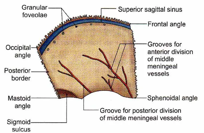
Internal Surface
It is highly concave, faces downward and medially, and is characterized by cerebral gyri impressions and grooves for meningeal vessels and venous sinuses.
The groove (or bony canal) extends upwards with a backward inclination from the inner surface of the sphenoidal (anterior-inferior) angle of the bone for the adjudication of the anterior (frontal) division of the middle meningeal vessels; the groove is created by the vein rather than the artery. One groove runs parallel to and about 1 cm behind the anterior margin, and it corresponds to the place of the precentral gyrus. Another group of grooves arises from the posterior portion of the inferior border and lodges the posterior (parietal) division of the middle meningeal vessels.
Along with the superior (sagittal) border is half of a groove that, when combined with a similar groove on the opposite parietal bone, forms a full sagittal sulcus for the accommodation of the superior sagittal sinus; the sagittal sulcus margins provide attachment to the dural fold, the falx cerebri. In old age, the sides of the sagittal sulcus have a variety of granular pits for the lodgement of arachnoid granulations, from which the cerebrospinal fluid is absorbed in the superior sagittal sinus.
Two imaginary lines can be drowned in the internal surface of the parietal bone for description purposes.
- A line stretches downward and forwards for about 7.5 cm from the superior margin, about 3 or 4 cm behind the anterosuperior angle. It refers to the central sulcus and divides the frontal lobe from the parietal lobe behind the brain.
- Another line, extending from the anterior inferior angle to the inner aspect of the parietal tuber, coincides with the posterior ramus of the lateral sulcus and connects the frontoparietal and occipitotemporal lobes above and in front, and the occipitotemporal lobes below and behind.
As a result, the parietal bone partly overlaps all four main lobes of the cerebral hemisphere. The parietal bone covers the following functional areas of the brain: Primary and secondary auditory regions, motor and somesthetic sensory areas, motor speech area (of Broca), and sensory speech area (of Wernicke) of the left hemisphere.
Superior Border
It has the longest serrated border and articulates with the opposite bone’s identical border at the sagittal suture.
Inferior Border
The anterior part of the border projects further below is beveled on the outside and articulates with the tip of the greater wing of the sphenoid bone in an overlapping manner. The intermediate and main parts are arched, beveled on the outside, and articulate with the upper border of the temporal bone’s squamous section. The inferior border’s posterior portion is serrated and articulates with the upper border of the mastoid part of the temporal bone.
The Anterior Border
It is serrated and beveled on the outer and inner surfaces in the upper and lower sections. It connects to the squamous aspect of the frontal bone and makes up half of the coronal suture.
The Posterior Border
It is dentated and forms half of the lambdoid suture by articulating with the superolateral border of the squamous aspect of the occipital bone.
The Anterosuperior Angle
It shapes the bregma, which is the meeting point of the coronal and sagittal sutures, by making a right-angle bend.
The Posterosuperior Angle(Occipital angle)
The lambda is formed by the union of the sagittal and lambdoid sutures.
The Anteroinferior Angle(Sphenoidal angle)
It juts downward and connects four bones separated by an H-shaped suture. Frontal bones are in front, squamous-temporal bones are behind, sphenoidal angle of parietal above, and tip of the greater wing of sphenoidal bones are below.
The pterion is the area occupied by the H-shaped suture, under which lies the anterior branch of the middle meningeal vessels and the stem of the lateral sulcus of the brain.
The Posteroinferior Angle(Mastoid angle)
The asterion is the meeting point of the parietal, occipital, and mastoid sections of the temporal bones.
Ossification of the Parietal Bone
The parietal bone ossifies in membrane from two primary centers in the parietal tuber that occur concurrently, one above the other, in the seventh week of intrauterine life. The centers fuse early to form a single center from which ossification spreads centrifugally, causing the borders to ossify before the angles. Fontanelles are the unossified membranous spaces at the four angles of the parietal bone. Six fontanelles involve both parietal bones in the neo-natal skull.
Growth
During the first two years, bone develops appositionally at the sutural membrane. As a result of the appositional formation, new layers of bones are deposited on the outer surface where tensile force is exerted by the expanding brain, while old layers are resorbed from the inner surface where compressive force is exerted. The growth processes of parietal bones can last up to seven or eight years and change the curvature of the cranial vault.
Clinical Points
Epidural Haematoma
The most common cause of epidural haematoma is a fracture of the parietal or temporal bone. The middle meningeal vessels, which lie in the bony grooves, may be torn, resulting in an arterial or venous haematoma. Arterial bleeding occurs quickly, and signs of brain compression usually appear within three hours. This is a medical emergency, and the cranial cavity should be explored right away to stop the bleeding and dissolve the blood clot.
In the case of an undiagnosed epidural haematoma caused by a head injury, symptoms include a brief loss of consciousness, accompanied by recovery from the original concussion, only to die hours later. This is referred to as the “talk-and-die syndrome.”
Osteoblastoma
An osteoblastoma is a rare form of primary bone tumor. It is exceedingly unusual to find it in the cranial vault.
Parietal Bone Summery
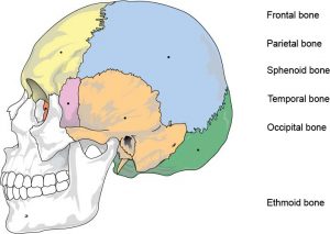
The parietal bones are bilateral skull bones that form the cranium’s superior and lateral walls. They are located above the parietal lobes of the brain and are only superficially protected by the epicranial aponeurosis. The parietal bones, along with the frontal, ethmoid, sphenoid, temporal, and occipital bones, comprise the neurocranium. The calvaria (skull cap) is formed by the majority of each parietal bone, while the remaining smaller portion is a component of the cranial base (basicranium). The cranium, and hence the parietal bones, serve to secure the underlying delicate brain.
The parietal bone is quadrilateral in shape and slightly curved. It is made up of two surfaces, four edges, and four angles. The borders shape various cranial sutures by articulating with neighboring skull bones. Numerous bony features and indentations on the surfaces refer to anatomical structures lying against the bone, such as superficial blood vessels of the brain.
Last Updated on February 23, 2022 by Learn From Doctor Team


