Table of Contents
Ethmoid Bone Overview
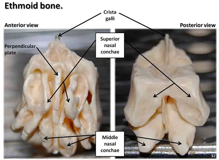
The ethmoid bone(Greek sierse) is a very light cuboidal bone located between the two orbits in the anterior of the base of the cranial cavity.
It forms;
- Part of the medial orbital walls
- Part of the nasal septum
- Part of the roof of the orbit
- And also the lateral walls of the nasal cavity
The ethmoid bone consists of;
- The cribriform plate
- The Perpendicular plate
- A pair of the labyrinth
Borders
Because of its central position inside the skull, the ethmoid bone comes into contact with 15 other skull bones. The most significant boundaries are:
- Anterior to the frontal bone
- Posterior with the sphenoid bone
- Inferior with the vomer and inferior nasal concha
The Cribriform Plate
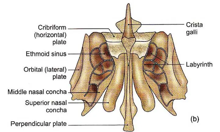
It is a horizontal perforated bony lamina that occupies the frontal bone’s ethmoidal notch. Foramina for olfactory nerve rootlets are present.
The Crista Galli
Crista Galli is a tooth-like upward projection in the anterior cranial fossa’s floor. The foramen transmitting the anterior ethmoidal nerve to the nasal cavity is located on the Crista Galli side.
The Perpendicular plate
It is a thin lamina that projects downward from the undersurface of the cribriform plate and forms the upper part of the nasal septum.
The Labyrinths
There are two light cubical masses suspended from the undersurface of the cribriform plate, one on either side of the perpendicular plate.
Each labyrinth also contains a large number of “air cells” that are divided into three groups: the anterior, middle, and posterior ethmoidal air sinuses.
Its surfaces are;
- To full anterior ethmoidal air cells, the anterior surface articulates with the frontal process of the maxilla.
- To full posterior ethmoidal air cells, the posterior surface articulates with the sphenoidal concha.
- Superior surface articulates with the frontal bone orbital plate.
- The inferior surface articulates with the maxillary nasal surface.
- The lateral surface serves as the orbit’s medial wall.
- The medial surface has a small superior nasal concha, a middle nasal concha, a superior meatus beneath the superior conchae, and a middle meatus beneath the middle concha.
Read The Parietal Bone
Deep Anatomy of the Ethmoid Bone
The ethmoid is a bone that is unpaired, fragile, pneumatic, and irregular. It is made up of a horizontal cribriform plate and two labyrinths that project downward from the lateral margin of the cribriform plates as a cuboidal, air-filled, and fragile bony unit.
The Cribriform Plate
It fills the ethmoidal notch between the 2 orbital plates of the frontal bone and has a series of apertures on every side for olfactory nerves with their arachnoid coverings to pass through. The plate has a posterior margin as well as upper and lower surfaces; the upper surface forms the floor of the anterior cranial fossa, and the lower surface forms the ceiling of the nasal cavity.
The posterior margin joins the ethmoidal spine of the sphenoid body.
The upper surface has a triangular raised crest, the crista galli, in the median plane. The crista galli has a long posterior border that connects to the anterior end of the falx cerebri; the crista’s short anterior border divides into two alae that connect to the frontal bone and form the posterior boundary of the foramen caecum. The perforated plate on either side of the crista galli supports the frontal lobe’s gyrus rectus as well as the olfactory bulb. The anterior portion of the upper surface on either side of the crista has a slit occupied by the dura mater and a narrow foramen for the passage of anterior ethmoidal vessels and nerves.
In the median plane, the lower surface has a quadrilateral, perpendicular plate that projects downward to form the upper part of the nasal septum. The anterior border of the perpendicular plate slopes downward and upward, articulating with the nasal spine of the frontal bone and the crest created by the joining of two nasal bones; the posterior border articulates with the sphenoidal crest in the upper part and the vomer in the lower part; and the inferior border is grooved to obtain the septal cartilage of the nose.
The Labyrinths
Each labyrinth is cuboidal in structure and comprises a number of ethmoidal air cells located between the orbital plate and the nasal plate. The ethmoidal sinuses are divided into three groups: anterior, middle, and posterior; in a disarticulated skull, the walls of certain air sinuses remain open and are only closed when adjacent bones are assembled for articulation. The sinuses, on the other hand, interact with the meatuses of the nasal cavity in the current state.
Each labyrinth’s upper surface articulates with the wide medial margin of the orbital plate of the frontal bone; anterior and posterior ethmoidal canals stretch transversely across the region of articulation and pass anterior and posterior ethmoidal vessels and nerves, meanwhile. The anterior surface of the maxilla articulates with the lacrimal bone and frontal process, completing the anterior ethmoidal sinuses. The posterior surface connects to the upper vertical portion of the sphenoidal concha as well as the orbital process of the palatine bone. The lower surface, represented by the orbital plate’s lower margin, articulates with the orbital surface of the body of the maxilla’s medial margin.
The orbital plate is a slim oblong plate that forms the orbit’s medial wall and overlaps the posterior and middle ethmoidal air cells.
The nasal plate on the labyrinth’s medial surface is part of the lateral wall of the nasal cavity. It terminates as a convexly curved disk, the middle nasal concha.
The latter is free in the intermediate part, but articulates in front with the ethmoidal crest of the frontal process of the maxilla and behind with the ethmoidal crest of the perpendicular plate of the palatine bone. The labyrinth’s middle meatus is a space lateral to the middle concha that has the following characteristics.
A hook-like uncinate process projects downward and backward and links with the inferior nasal concha’s ethmoidal process.
The bulla ethmoidalis is a rounded elevation that extends into the lateral wall of the middle meatus and contains middle ethmoidal air cells; middle ethmoidal cells open on or above the bulla.
The semilunar hiatus is a space between the uncinate process below and in front of the uncinate process and the bulla ethmoidalis above and behind it. The infundibulum is a bony passage that stretches upward and forwards inside the labyrinth from the anterior part of the hiatus semilunaris, receiving the opening of anterior ethmoidal air cells and communicating with the frontal sinus through the frontonasal duct.
The superior meatus is a small oblique groove on the nasal surface above the middle nasal concha, which is confined above by a curved plate known as the superior nasal concha; the superior meatus receives the openings of the posterior ethmoidal sinuses. The spheno-ethmoidal recess, which is located above the superior concha and between the ethmoid and the body of the sphenoid, is where the sphenoidal sinus opens.
The Ossification
The ethmoid bone is formed by the ossification of three cartilaginous nasal capsule centers: one for each labyrinth and one for the perpendicular layer.
The labyrinth centers appear during the fourth and fifth foetal months, and the perpendicular plate centers appear during the first year. In the second year, the three elements are fused. In intrauterine life, ethmoidal air cells form as narrow pouches.
Clinical Points
Fracture of the Cribriform Plate
A cribriform plate fracture may cause rhinorrhea and the discharge of cerebrospinal fluid through the nose.
Fracture of the Ethmoidal Labyrinth
Crushing injuries to the ethmoidal labyrinth cause disruptions in the separation between the orbital and nasal cavities, resulting in exophthalmos when blowing through the nose. This may be due to the spread of air bubbles in the subcutaneous tissue of the eyelids and face, resulting in surgical emphysema.
Ethmoidal Sinusitis
The ethmoid sinuses are hollow spaces in the nasal bones. They have a mucus coating to keep the nose from drying out. The ethmoid sinuses may become inflamed, causing pressure and discomfort around the nose and between the eyes.
Summary of the Ethmoid Bone
The ethmoid bone is a single porous bone that forms the midfacial region of the skull and forms the middle area of the viscerocranium. It aids in the formation of the orbit, nasal cavity, nasal septum, and anterior cranial fossa floor.
The ethmoid bone, which develops through the process of endochondral ossification, is an essential part of your skull, particularly because of the cribriform plate, which enables olfactory fibers to move through so you can smell stuff.
Last Updated on February 23, 2022 by Learn From Doctor Team


