Table of Contents
Sphenoid Bone Overview
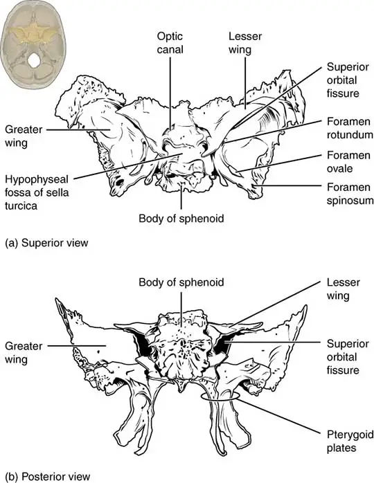
The sphenoid bone(Greek wedge) bone is shaped like a bat with wings outstretched. It consists of the following parts;
- A body in the center of the bone
- Two lesser wings from the anterior part of the body
- Two greater wings from the lateral part of the body
- Two downward-directed pterygoid (wing-like) processes from the junction of the body and greater wings.
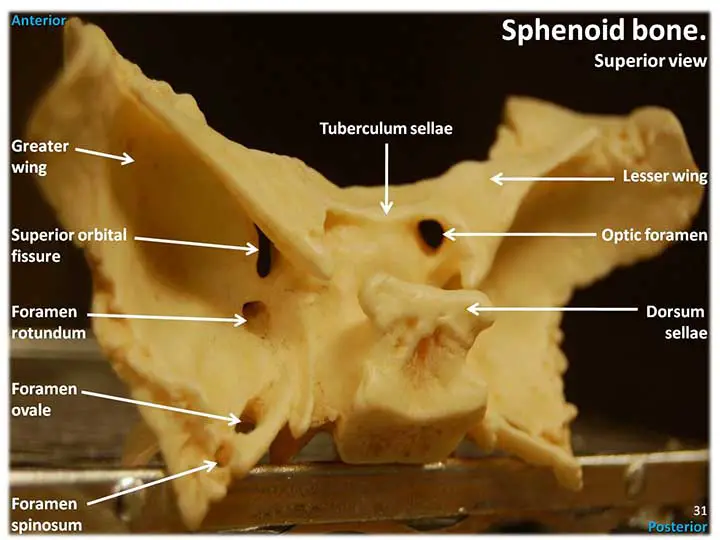
Body of the Sphenoid
It has six surfaces and encloses a pair of sphenoidal air sinuses.
The surface
Superior or Cerebral Surface
It articulates with the ethmoid bone anteriorly and the basilar part of the occipital bone posteriorly. It presents;
- The jugum sphenoidale
- The sulcus chiasmaticus
- The tuberculum sellae
- The sella turcica
- The dorsum sellae
- The clivus
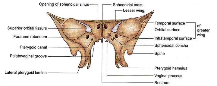
The Inferior Surface
- The rostrum of the sphenoid
- The sphenoid conchae
- The vaginal processes of the medial pterygoid plate
Anterior Surface
The sphenoidal crest articulates with the perpendicular plate of the ethmoid to form a small part of the nasal septum.
The opening of the sphenoidal air sinus is visible.
The sphenoidal conchae close the sphenoid air sinuses while leaving the openings open. Each half of the anterior surface is divided into two sections: superolateral and inferomedial.
To complete the posterior ethmoidal air sinuses, the superolateral depression articulates with the ethmoid labyrinth.
The posterior part of the nose’s root is formed by the inferomedial smooth triangular area.
Posterior Surface
It connects to the basilar part of the occipital bone.
Lateral Surface
Carotid sulcus, a large groove shaped like the letter ‘f’ for cavernous sinus and internal carotid artery lodging. Gelow sulcus articulates laterally with the greater wing of the sphenoid, also with the pterygoid process, which is directed downwards.
Sphenoidal Air Sinuses
There are asymmetrical air sinuses in the sphenoid’s body that are closed by sphenoidal conchae. In the sphenoethmoidal recess above the superior concha, the sinus opens in the lateral wall of the nose.
Greater Wings of the Sphenoid
These are two prominent Processes that curve laterally and upwards from the body’s sides. It has three different surfaces.
Superior or Cerebral Surface
The floor of the middle cranial fossa is formed by this surface and it presents;
Before backward:
- The foramen rotundum
- The foramen ovale
- The emissary sphenoidale foramen
- The foramen spinosum
Lateral Surface
The infratemporal crest, a horizontal ridge, divides this surface into an upper or temporal surface and a lower or infratemporal surface. It has foramen ovale and foramen spinosum’s piercings. Its posterior section displays the sphenoid spine.
Orbital Surface
Forms the posterior wall of the orbit’s lateral wall.
It has a small tubercle on its medial border for the attachment of a common tendinous ring for the origin of the recti muscles of the eyeball. The grooved region forms the posterior wall of the pterygopalatine fossa and is penetrated by the foramen rotundum below the medial end of the superior orbital fissure.
Borders cover the greater wing of the sphenoid.
Lesser Wings of the Sphenoid
Lesser wings are two triangular plates that project laterally from the body’s anterosuperior region. It consists of the following elements;
- The base forms the wing’s medial end. It is attached to the body through two roots that encircle the optic canal.
- The wing’s lateral end is formed by the tip.
- The superior surface forming the anterior cranial fossa’s floor
- The superior orbital fissure’s inferior surface forms the upper boundary.
- The anterior border articulates with the posterior border of the frontal bone’s orbital plate.
- The posterior border of the brain is free and projects into the stem of the lateral sulcus. It terminates medially in the anterior clinoid process.
The Superior Orbital Fissure
It is a triangular gap that connects the middle cranial fossa to the orbit. The structures that pass through it are added to the list of foramina and structures that pass through them.
The Pterygoid Process
One pterygoid (Greek wing) process projects downwards from the body’s junction with the greater wing of the sphenoid on either side.
Each pterygoid process is divided into medial and lateral pterygoid plates. The plates are fused in the upper parts but divided into the lower parts by the pterygoid fissure. The pterygoid plates surround a “V-shaped interval” called the pterygoid fossa posteriorly. The medial pterygoid plate has a scaphoid fossa in its upper half.
Borders of the Sphenoid
The sphenoid bone shares a boundary with the frontal bone (via the sphenofrontal suture), the parietal bone (via the sphenoparietal suture), the temporal squamous portion (via the sphenosquamosal suture), and the occipital bone (via the spheno-occipital suture). The spheno-occipital suture disappears by the age of 25 when the sphenoid and occipital bones fuse during puberty (“tribasilar bone”).
Deep Anatomy of the Sphenoid Bone
The most complicated bone in the human body is the sphenoid bone. It is also recognized as the “wasp bone” due to its shape. It occupies the majority of the middle portion of the base of the skull and extends to the floor of the middle cranial fossa.
This bone is connected with and has strong links to soft tissue structures such as the cranial nerves and portions of the brain. The sphenoid bone is also involved in the formation of many of the cranium’s foramina and canals.
Ossification
The sphenoid bone’s body and lesser wings mature via classic endochondral ossification, while the pterygoid processes mature via intramembranous ossification.
The formation of the greater wings of the sphenoid is unique because they are the only bony elements of the skull that undergo both endochondral and intramembranous ossification.
Openings
This cranial bone has many openings that allow nerves and blood vessels to pass into and out of the cranial cavity. The optic canal, superior orbital fissure, foramen rotundum, foramen lacerum, foramen spinosum, and foramen ovale are among them. The foramen lacerum is located between the sphenoid, the basilar portion of the occipital, and the temporal bone’s apex, and it also includes connective tissue.
Functions
The sphenoid bone serves a variety of important roles. It, along with the orbital floor, contributes to the formation of the base and lateral sides of the skull. The skull’s multiple articulations with other bones lend it rigidity. It serves as an attachment point for many masticatory muscles. The facial skeleton is formed by a portion of the sphenoid. In addition, the sphenoid has many fissures and foramina that carry blood vessels and nerves from the skull to the head and neck. The sphenoid’s body contains a cavity with a sinus that interacts with the nasal cavity. These sphenoidal sinuses help to lighten the skull and increase resonance.
Blood Supply, Nerve Supply, and Lymphatics Drainage
The blood vessels move into the foramina in the sphenoid bone’s wings. The ophthalmic artery enters the orbit through the optic canal. The ophthalmic veins exit the orbit through the superior orbital fissure. The accessory meningeal artery enters the skull through the foramen ovale. The foramen spinosum allows the middle meningeal artery and vein to pass through. Only a few small meningeal arterial branches and veins pass through the foramen lacerum, which can be closed off by cartilage.
Several nerves travel through the sphenoid bone’s foramina, fissures, and canal. The optic canal enables the optic nerve to pass through (CN II). The superior orbital fissure carries the superior and inferior divisions of the oculomotor nerve (CN III), trochlear nerve (CN IV), ophthalmic nerve (CN V1), abducens nerve (CN VI), and sympathetic fibers from the cavernous plexus. The maxillary division of the trigeminal nerve is contained within the foramen rotundum (CN V2). The foramen ovale provides access to the trigeminal nerve’s mandibular division (CN V3), the motor root of the trigeminal nerve, and the lesser petrosal nerve. The meningeal branch of the mandibular nerve passes through the foramen spinosum.
Clinical Points
Sphenoid Fracture
Sphenoid fractures occur as a result of damage to the orbit or the base of the skull. Since the sphenoid is closely associated with numerous nerves that pass through it, these fractures can result in multiple neurological sequelae. These fractures are linked to vision loss and ocular damage. Among the signs and symptoms are the Battle sign, hemotympanum, and cranial nerve palsies.
Wing Dysplasia
Sphenoid wing dysplasia in patients with neurofibromatosis type 1 can cause difficult and severe changes, including vision loss.
Surgical Point
In cases of pituitary tumors that need surgical removal, trans-sphenoidal surgery is the treatment of choice. The pituitary gland is accessed by moving surgical instruments through the nose and sphenoidal sinuses to enter the sella turcica. This trans-sphenoidal technique could also be used to gain entry to structures other than the pituitary, such as a craniopharyngioma.
Sinusitis (Sphenoidal)
Infections of the sphenoid sinuses can cause acute and chronic sinusitis. The sphenoid sinuses are rarely involved in isolation, commonly occurring in conjunction with frontal and ethmoidal sinusitis. Fever, fatigue, and a post-nasal drip are all symptoms. If not treated immediately, it can lead to life-threatening complications such as meningitis, brain abscess, and cranial nerve involvement.
Sphenoid Bone Summery
The sphenoid is one of the twenty-two bones that make up the skull and serves to bind the neurocranium to the facial skeleton. It is a single bone in the cranial cavity located posterior to the frontal bone but anterior to the occipital. It gets its name from the Greek word sphenoeides, which means “wedge-shaped.” It has many foramina and fissures on its surface that enable nerves and blood vessels to enter and leave the cranial cavity. It has a central body and two lateral wings on either side, similar to a butterfly or a bat. It is one of the orbital bones, forming the posterior surface in particular. The sphenoid articulates with the frontal, parietal, ethmoid, zygomatic, temporal, occipital, palatine, and vomer bones. The relationship between the sphenoid, ethmoid, and frontal bone may have many morphological variations. Together, these give the skull rigidity and stability, making it a perfect place to house the brain.
Last Updated on February 23, 2022 by Learn From Doctor Team


