Table of Contents
Brachiocephalic Vein(Innominate Vein) Overview
The brachiocephalic veins are broad paired valveless asymmetric veins that drain the head, neck, upper limbs, and a portion of the thorax and mediastinum.
Origin
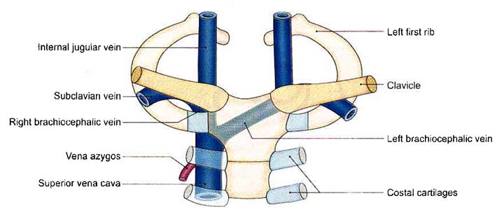
The internal jugular and subclavian veins join at the base of the neck to form the brachiocephalic veins, which are located posterior to the medial ends of the clavicles.
Course
The left brachiocephalic vein is about 6 cm long and runs an oblique path to the right through the superior mediastinum anterior to the branches of the aortic arch to join the right brachiocephalic vein posterior to the first sternocostal joint to form the superior vena cava.
The right brachiocephalic vein is much narrower, measuring around 2.5 cm in length and running vertically anterior to the brachiocephalic trunk. As it is joined from the left by the left brachiocephalic vein, it becomes the superior vena cava.
Tributaries
Left Brachiocephalic Vein
- The left vertebral vein
- The left inferior thyroid vein
- The left internal thoracic vein
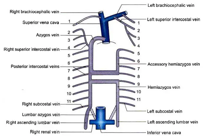
- The left supreme intercostal vein
- The left superior intercostal vein
- The thymic veins
- The pericardiophrenic veins
Right Brachiocephalic Vein
- The right vertebral vein
- The right inferior thyroid vein
- The right internal thoracic vein
- The right supreme intercostal vein
The superior intercostal vein on the right normally drains into the azygos vein.
Relation
Anterior
The pleura, upper lobes, thymus, manubrium
Posterior
The aortic arch (left brachiocephalic vein), great vessels, the dome of the pleura
Read The Brachiocephalic Trunk
Detailed Anatomy of the Brachiocephalic Vein
The brachiocephalic vein, also known as the innominate vein, is a paired superior mediastinum vein that drains venous blood from the head and neck, upper limbs, and upper thorax. The internal jugular and subclavian veins confluence on either side, just posterior to the sternoclavicular joint, to form it.
The superior vena cava is formed when the left and right brachiocephalic veins join at the level of the inferior border of the first right costal cartilage.
Beginning, Course, and Termination
The left and right brachiocephalic veins arise from the union of the internal jugular and subclavian veins and begin posteriorly to the respective left and right sternoclavicular joints. This union is known as the venous angle, and it is where the thoracic duct on the left and the right lymphatic trunk on the right drain lymph into the venous circulation. As a result, the brachiocephalic vein is thought to be the point of convergence of the venous and lymphatic systems.
The left brachiocephalic vein runs obliquely downwards and medially and is about 6 to 8 centimeters long. The left brachiocephalic vein is connected to the trachea, the left phrenic and vagus nerves, the left internal thoracic artery, and the three main branches of the arch of the aorta: the brachiocephalic trunk, the common carotid artery, and the left subclavian artery. The sternohyoid and sternothyroid muscles distinguish the sternoclavicular joint from the left brachiocephalic vein, which is in contact with and partially embedded in the thymus gland. The left brachiocephalic vein begins adjacent to the medial surface of the apex of the left lung and is eventually overlapping by the right pleura.
The right brachiocephalic vein is approximately 2 centimeters long and has a shorter and more vertical path. It connects to the brachiocephalic trunk and the right vagus nerve anterolaterally. The right brachiocephalic vein is located anterior to the right pleura, phrenic nerve, and internal thoracic artery in its initial path, but later turns to its medial side.
The left and right brachiocephalic veins converge at the level of the inferior border of the 1st right costal cartilage to form the superior vena cava, which drains into the right atrium of the heart.
Tributaries and Drainage of the Vein
The tributaries to the left and right brachiocephalic veins are unique. The brachiocephalic veins receive venous blood from the head, neck, upper limb, and upper part of the thorax through these tributaries.
Note: Read the tributaries in the Overview part above for a better understanding.
The Right Brachiocephalic Vein
The right vertebral, internal thoracic, and inferior thyroid veins, as well as the right posterior intercostal vein of the 1st intercostal space, are tributaries of the right brachiocephalic vein.
The Left Brachiocephalic Vein
The left vertebral, internal thoracic, inferior thyroid, and superior intercostal veins are tributaries of the left brachiocephalic vein. Furthermore, it also receives the 1st intercostal space’s thymic, supreme intercostal, pericardiacophrenic, and left posterior intercostal veins.
Embryology
The cardinal venous system gives rise to the brachiocephalic veins. This system, which consists of three pairs of cardinal veins, starts to drain the embryo’s body in the fifth week of life. The right brachiocephalic vein arises from the right anterior cardinal vein, the right normal cardinal vein, and the right horn of the sinus venosus. The left anterior cardinal vein gives rise to the left brachiocephalic vein.
The mesoderm is the leaflet of embryological derivation.
Clinical Points
The Retro-aortic Left Brachiocephalic Vein
The retro-aortic left brachiocephalic vein is an unusual vascular variant in which the left brachiocephalic vein runs more inferiorly through the superior mediastinum, inferior to the aortic arch and posterior to the ascending aorta before joining the right brachiocephalic vein to form the superior vena cava. The vein typically passes higher and more horizontally through the superior mediastinum, anterior to the branches of the aortic arch.
Brachiocephalic Vein Thrombosis
Brachiocephalic vein thrombosis accounts for a small of cases of deep vein thrombosis. The prevalence appears to be increasing, particularly because of increased use of indwelling central venous catheters.1,2 proximal upper extremity deep vein thrombosis is defined as thrombosis involving the axillary or more proximal deep veins, and distal upper extremity deep vein thrombosis is defined as thrombosis of the brachial or more distal deep arm veins. Axillary and subclavian veins are most frequently affected.
Brachiocephalic Vein Aneurysm
Just 36 cases of brachiocephalic vein aneurysms have been identified in the literature. They usually manifest as mediastinal widening on chest X-ray, with thromboembolism or mass impact on adjacent structures, or rupture. While imaging is normally sufficient to recognize and describe an aneurysm, some diagnostic pitfalls may lead to misinterpretation and misdiagnosis. Often exploratory surgery is required to confirm a diagnosis. Brachiocephalic vein aneurysms have been treated conservatively in the previous time with watchful waiting, antithrombotic treatment, or anticoagulation, as well as surgically, depending on the patient’s presentation and aneurysm characteristics. Endovascular therapy is now becoming a viable therapeutic alternative. Following surgical surgery, the prognosis is excellent, with no confirmed cases of recurrence.
Lipoma in the Brachiocephalic Vein
Intravascular lipomas are uncommon primary venous tumors that develop from vein walls, most usually from the inferior vena cava, and occur in 0.35 percent to 0.5 percent of all CT scans. They occur less often from the right brachiocephalic vein, superior vena cava, iliac, and femoral veins.
Brachiocephalic Vein Summery
The brachiocephalic veins, also known as the innominate veins, are massive venous structures that arise in the thorax from the union of the subclavian vein and the internal jugular vein. The superior vena cava is formed by the union of the left and right brachiocephalic veins on the right side of the upper chest. These vessels are an important part of the human circulatory system because they aid in the drainage of deoxygenated blood from the head and upper limbs.
Last Updated on February 23, 2022 by Learn From Doctor Team


